


Home / Training / Manuals / Atlas of breast cancer early detection / Cases
Atlas of breast cancer early detection
Go back to the list of case studies
.png) Click on the pictures to magnify and display the legends
Click on the pictures to magnify and display the legends
BI-RADS Category (at follow-up (2018)): 2 (benign)
| Case number: | 168 |
| Age: | 65 |
| Clinical presentation: | Postmenopausal woman with average risk of developing breast carcinoma presented for screening. CBE revealed a small nodule palpable in the lower quadrant of the left breast. |
Mammography:
 |  |
| Breast composition: | ACR category a (the breasts are almost entirely fatty) | Mammography features: |
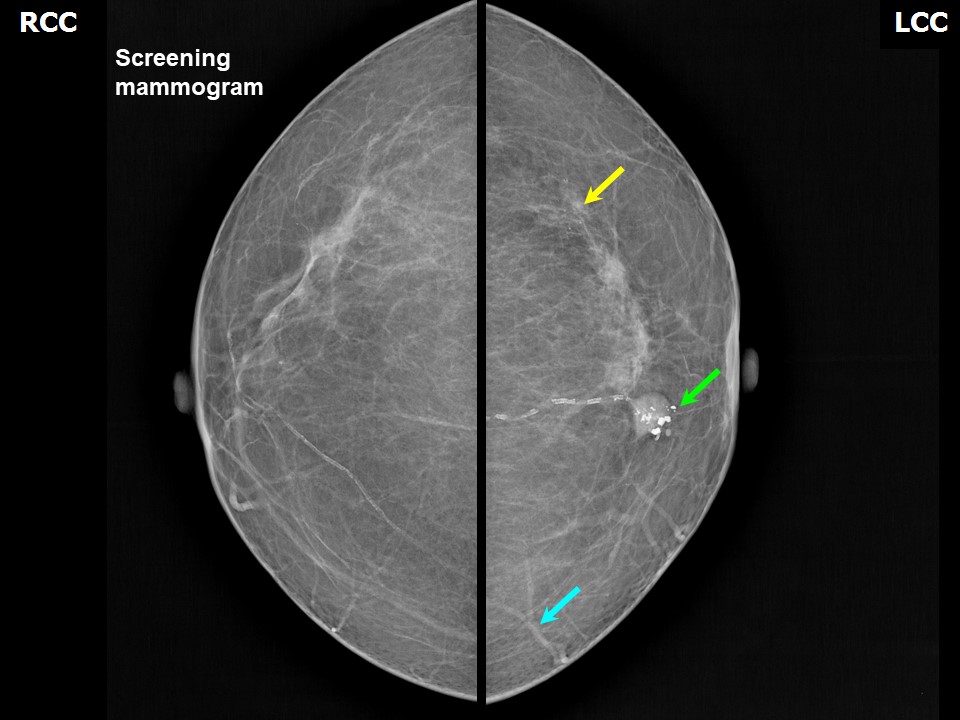
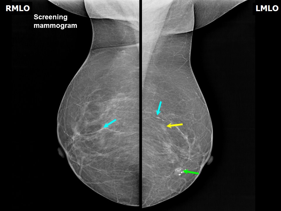
| ‣ Location of the lesion: | 2017: Right breast, vascular calcification |
| ‣ Mass: | |
| • Number: | 0 |
| • Size: | |
| • Shape: | None |
| • Margins: | None |
| • Density: | None |
| ‣ Calcifications: | |
| • Typically benign: | Vascular calcification |
| • Suspicious: | None |
| • Distribution: | None |
| ‣ Architectural distortion: | None |
| ‣ Asymmetry: | None |
| ‣ Intramammary node: | None |
| ‣ Skin lesion: | None |
| ‣ Solitary dilated duct: | None |
| ‣ Associated features: | None |
| Breast composition: | ACR category a (the breasts are almost entirely fatty) | Mammography features: |
| ‣ Location of the lesion: | 2017: Left breast, lower inner quadrant at 7 oclock, middle third |
| ‣ Mass: | |
| • Number: | 1 |
| • Size: | 1.6 × 1.2 cm |
| • Shape: | Oval |
| • Margins: | Circumscribed with perilesional halo |
| • Density: | Equal |
| ‣ Calcifications: | |
| • Typically benign: | Coarse |
| • Suspicious: | None |
| • Distribution: | Macrocalcifications within the mass |
| ‣ Architectural distortion: | None |
| ‣ Asymmetry: | None |
| ‣ Intramammary node: | None |
| ‣ Skin lesion: | None |
| ‣ Solitary dilated duct: | None |
| ‣ Associated features: | Calcifications |
| Breast composition: | ACR category a (the breasts are almost entirely fatty) | Mammography features: |
| ‣ Location of the lesion: | 2017: Left breast, upper outer quadrant at 12 oclock, middle third |
| ‣ Mass: | |
| • Number: | 0 |
| • Size: | |
| • Shape: | None |
| • Margins: | None |
| • Density: | None |
| ‣ Calcifications: | |
| • Typically benign: | None |
| • Suspicious: | Suspicious calcifications |
| • Distribution: | Numerous calcifications, > 5 in number, seen grouped together, in less than 2 cm of breast tissue, regional distribution |
| ‣ Architectural distortion: | None |
| ‣ Asymmetry: | None |
| ‣ Intramammary node: | None |
| ‣ Skin lesion: | None |
| ‣ Solitary dilated duct: | None |
| ‣ Associated features: | None |
 |  |
 |
| Breast composition: | ACR category a (the breasts are almost entirely fatty) | Mammography features: |
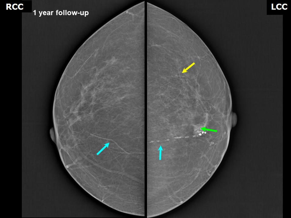
| ‣ Location of the lesion: | 2018: Follow-up mammogram shows stable calcifications in the upper outer quadrant of the left breast, Middle third, BI-RADS 2 (benign) |
| ‣ Mass: | |
| • Number: | 0 |
| • Size: | |
| • Shape: | None |
| • Margins: | |
| • Density: | |
| ‣ Calcifications: | |
| • Typically benign: | None |
| • Suspicious: | None |
| • Distribution: | |
| ‣ Architectural distortion: | |
| ‣ Asymmetry: | |
| ‣ Intramammary node: | |
| ‣ Skin lesion: | |
| ‣ Solitary dilated duct: | |
| ‣ Associated features: | None |
BI-RADS:
BI-RADS Category (2017): 3 (probably benign)BI-RADS Category (at follow-up (2018)): 2 (benign)
Case summary:
| Postmenopausal woman presented with left breast nodule. Diagnosed as involuting (partially calcified) fibroadenoma in the left breast, BI-RADS 2 on Imaging. Survey of bilateral breast mammogram reveals grouped calcifications of suspicious morphology in the left breast on initial assessment, BI-RADS 3. Regular annual follow-up reveals stable unchanged calcifications in the left breast, diagnosed as BI-RADS 2 on imaging. |
Learning points:
|